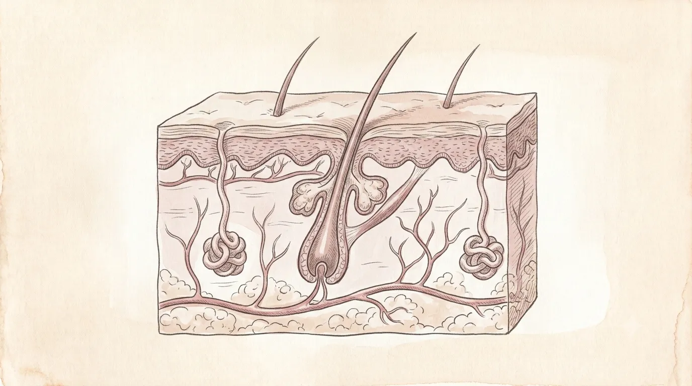
A single square centimetre can hold hundreds of sweat glands and metres of blood vessels.
Integumentary System
Skin
IntegumentumThe living boundary
Protects, senses, and cools
The body’s largest organ — a living barrier that senses touch, holds in water, and regulates temperature.
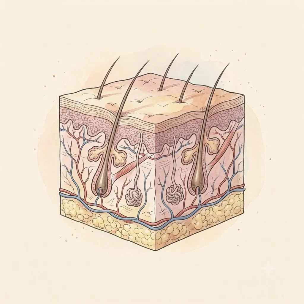
- Stratum CorneumDead cells forming the real barrier
- EpidermisOuter protective layer
- DermisNerves, vessels & glands
- Hair FollicleAnchors each hair
- Sebaceous GlandOil that waterproofs skin and hair
- Sweat GlandEvaporative cooling — the body's thermostat
- MelanocytesPigment cells shielding DNA from ultraviolet light
- Capillary LoopDermal vessels that flush or clamp to manage heat
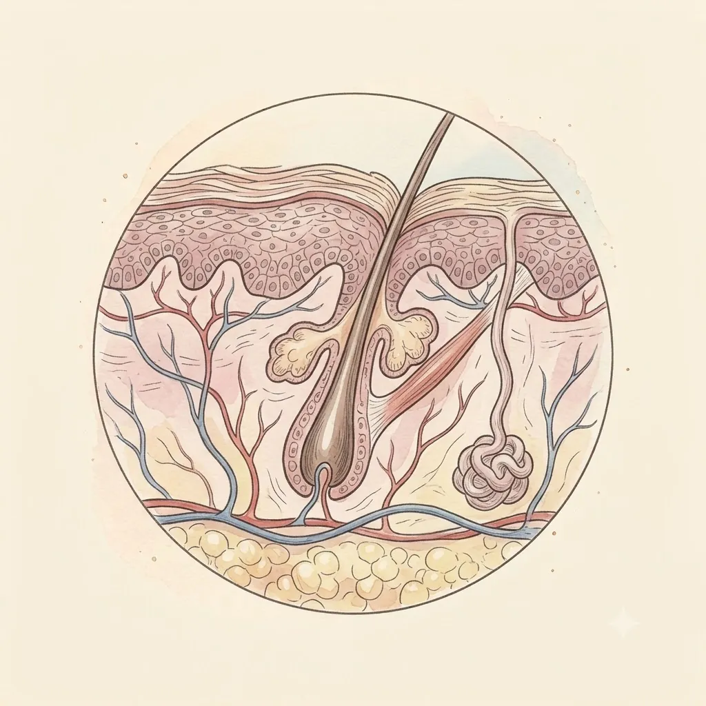 Under the lens
- Nerve EndingsTouch, temperature, and pain receptors
- HypodermisFat and insulation
- Stratum BasaleThe germinal row — every keratinocyte is born here, and the melanocytes sit among them
- Arrector PiliSmooth muscle from follicle to dermis; sympathetic fibres contract it and the hair stands up
- Hair BulbThe growing end, where matrix cells divide around a dermal papilla that feeds them
- Reticular LayerThe deep dermis — coarse collagen bundles and elastic fibres that give skin its strength
- Deep Vascular PlexusArtery and vein at the dermal-fat border, the source of everything above
- Skin LigamentsFibrous strands crossing the fat, tying the dermis down to the deep fascia
Structure
- Stratum corneumDead flattened cells in a lipid mortar — the actual waterproof barrier. Only fifteen or twenty cells deep over most of the body, but many times that on the palms and soles, where a further clear layer is packed in beneath it
- EpidermisLiving layers below, pushing new cells upward over about four weeks
- Basement membraneThe glue between epidermis and dermis; it fails in blistering disease
- DermisCollagen and elastin, holding vessels, nerves, glands, and follicles
- HypodermisFat for insulation, padding, and energy storage — the superficial fascia, split over the abdomen into a fatty layer and a membranous one, and crossed by the skin ligaments that anchor the dermis to the deep fascia below
- Sits
- Covering the entire body
- Size
- About 2 m² spread flat
- Weight
- 3.5–5 kg
The lesson
Five layers stand between the body and the world, from the dead cells of the stratum corneum that do the actual waterproofing, down through the living epidermis and collagen-rich dermis, to the fatty hypodermis beneath. It renews from the bottom up — new cells push older ones outward over about four weeks until they are shed.
The barrier is made of dead cells, on purpose
The waterproof layer is not living tissue. A cell born at the base spends its life climbing, and on the way it fills with keratin, dismantles its own nucleus and dies in a controlled sequence. The barrier is what it leaves behind, not what it was.
Dying is the last step of the job rather than a failure of it.
What actually stops water is the arrangement. Flattened dead cells stack like bricks, and as they go the cells extrude a lipid mortar that fills every gap between them. Neither alone would hold — it is the composite that works.
Strip the lipid with harsh soap, a solvent or a long hot wash and the bricks stay exactly where they were while the barrier leaks, which is why skin that looks perfectly intact can still be dry and irritable.
Remember
- Keratinocytes die in a programmed way; the dead layer is the working barrier
- Bricks and mortar — flattened cells set in extruded lipid
- Removing the lipid breaks the barrier without visibly damaging the skin
It keeps water in more than it keeps things out
The harder job faces inward. A land animal’s standing threat is losing water to dry air, and skin’s water-holding is what makes life out of water possible at all.
The scale of that job only becomes obvious when it fails: extensive burns kill largely through fluid loss and through infection entering the opening, not through the injury itself.
The same sheet runs the body’s temperature. Its blood supply is far larger than the tissue’s own metabolism could ever need, because it is a radiator rather than a supply line — opened wide to dump heat, shut down to hoard it. Sweat then costs heat to evaporate and takes that heat from the skin.
Flushing and pallor are that thermostat, visible from outside.
Remember
- Holding water in is the primary job; keeping things out is secondary
- Dermal blood flow is sized for heat exchange, not for feeding the tissue
- Burns are dangerous chiefly through fluid loss and infection
Several different senses in one sheet
Touch is not one sense. The dermis holds distinct endings answering to different things — some fire only at the moment pressure changes, others keep firing for as long as it lasts, others report stretch, vibration, temperature or damage.
What arrives as a single impression is several channels being read together.
The endings are not spread evenly, and the brain assigns cortex in proportion to their density rather than to area. A fingertip packs them close and commands a large share of the sensory map; a stretch of back carries few and commands very little.
Two points a centimetre apart feel like two on a fingertip and like one on the back — the same skin, sampled at completely different resolutions.
Remember
- Separate receptors for changing pressure, sustained pressure, vibration, temperature and damage
- Receptor density varies enormously from one part of the body to another
- Cortical territory follows density, not surface area
Clinical use
Sheds around 500 million cells
Three layers — epidermis, dermis, and hypodermis — each with a distinct job.
Always read a plate alongside the clinical picture.