The stapedius, barely a centimetre long and hidden in the middle ear, is the smallest skeletal muscle in the body; the gluteus maximus, at the opposite end of the skeleton, is the largest and among the most powerful.
All modules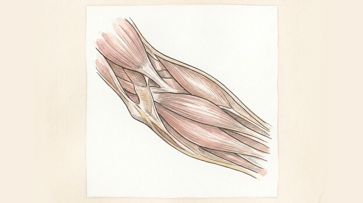 Musculoskeletal System
Musculoskeletal SystemMuscles: the lesson
Structure and story of the muscles, layer by layer.
Musculoskeletal System
Muscles
Systema musculareThe engine wrapped around the frame
Moves the skeleton, holds every joint stable against gravity, generates most of the body's resting heat, and powers breathing, swallowing and speech
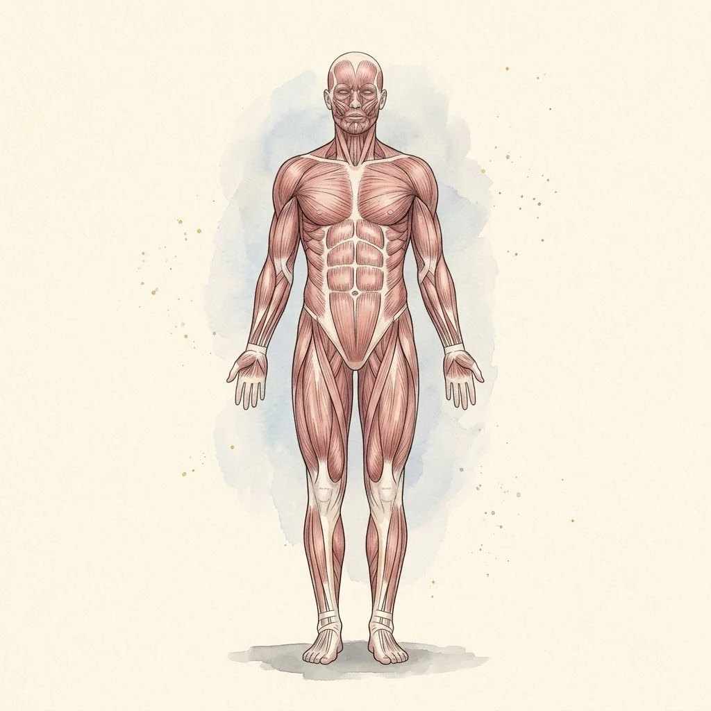
About 640 skeletal muscles — the body's only source of movement, each one a bundle of fibres that shortens on a nerve's command and lets go the instant the command stops.
- DeltoidThe rounded cap of the shoulder, its three parts able to lift the arm in almost any direction; the standard site for an intramuscular injection
- Biceps BrachiiTwo heads crossing both the shoulder and the elbow, flexing the elbow and turning the forearm to face the palm up
- Pectoralis MajorThe broad fan of the chest wall, drawing the arm across the body and forming the anterior wall of the armpit
- Rectus AbdominisThe paired vertical strap of the abdominal wall, its tendinous intersections giving a lean abdomen its visible segments
- Gluteus MaximusThe largest and most powerful muscle in the body, driving the hip from a bent to a straight position in climbing and sprinting
- GastrocnemiusThe calf's two-headed muscle, crossing both knee and ankle to point the foot and power the push-off of every stride
- TrapeziusA broad sheet from skull to mid-back that lifts, rotates and steadies the shoulder blade against the trunk
- Latissimus DorsiThe widest muscle of the back, pulling the raised arm down and back — the muscle a pull-up is built on
 Under the lens
- Triceps BrachiiThe elbow's sole extensor, three heads converging on one tendon at the point of the elbow
- External ObliqueThe outermost of the abdominal wall's three angled sheets, rotating the trunk and bracing it against the muscle beneath
- Quadriceps FemorisFour heads sharing one tendon around the patella, the body's principal knee extensor and the muscle that fails first climbing stairs
- HamstringsThree muscles down the back of the thigh, extending the hip and flexing the knee — and among the most commonly strained in sprinting
- Tibialis AnteriorThe shin's prominent tendon, lifting the front of the foot clear of the ground during the swing of each step
- MasseterThe strongest muscle for its size in the body, closing the jaw with a bite force that can exceed body weight
- SternocleidomastoidThe cord that stands out turning the head to one side, running from behind the ear to the collarbone and breastbone
- DiaphragmThe dome of muscle separating chest from abdomen; its flattening on contraction is what actually draws air into the lungs
Structure
- EpimysiumThe tough outer sheath wrapping the whole muscle, continuous with the tendon at each end and with the deep fascia around it
- FascicleA visible bundle of muscle fibres — the grain you see when meat is cut, and the unit the muscle's power and pull direction are built from
- PerimysiumThe connective-tissue wrap around each fascicle, carrying the branch of the muscle's artery and nerve that supplies it
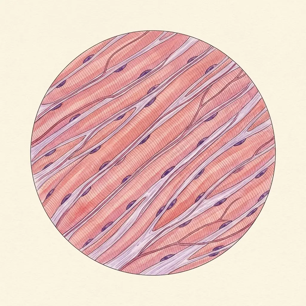
- Muscle fibreA single, multinucleated cell running the fascicle's length, packed with the sarcomeres that do the actual contracting
- EndomysiumThe thinnest wrap of all, around one fibre alone, holding the capillaries and nerve endings each fibre depends on directly
- Sits
- Wrapped around every bone the skeleton provides, from the scalp to the sole of the foot
- Size
- Around 640 named muscles, most of them paired left and right
- Weight
- About 40% of body weight in a lean adult — the single largest tissue in the body
The lesson
A muscle looks like dead meat on a plate and is the opposite of that: a living cable of thousands of fibres that converts a nerve's electrical signal into force by a molecular ratchet, arranged so that muscles only ever work in opposing pairs, and fuelled by three different chemical systems depending on how hard and how long the work has to last.
Inside every fibre is a molecular ratchet
A muscle fibre is packed with sarcomeres end to end, and a sarcomere is where the actual shortening happens: thin filaments of actin and thick filaments of myosin overlap, and myosin's heads reach out, grip actin, pull, release, and reach again — a ratchet stroke repeated thousands of times a second across the whole fibre.
Neither filament changes length; the sarcomere shortens because the two slide further over each other, which is why the whole theory is named for sliding filaments rather than for shrinking ones.
The ratchet is switched on by calcium.
An action potential travelling into the fibre releases calcium stored in the sarcoplasmic reticulum, which binds troponin and swings tropomyosin off the site myosin needs to grip — remove the nerve signal and the calcium is pumped back inside within milliseconds, tropomyosin covers the site again, and the fibre relaxes.
Every stroke of the ratchet also burns ATP, which is what actually lets go of the myosin-actin bond; without it the bond locks instead of releasing, which is exactly what happens throughout the body after death, and is why rigor mortis sets muscle rigid until the fibres themselves begin to break down.
Remember
- Actin and myosin filaments slide over each other; neither filament shortens itself
- Calcium released by a nerve signal exposes the site myosin needs to grip
- ATP is spent to release the grip, not to create it — its absence is why rigor mortis locks muscle rigid
A muscle can only pull — so everything comes in pairs
Muscle has exactly one trick: it shortens and pulls. It cannot push a joint back the other way, so every mobile joint is crossed by at least one opposing pair — an agonist doing the movement and an antagonist that lengthens to allow it and then reverses the job to bring the joint back.
The biceps flexes the elbow, the triceps extends it; contract both at once instead and the joint locks rigid rather than moving, which is exactly the trick a joint under threat of injury uses to protect itself.
Most real movements need more than one pair.
Synergists assist the prime mover or fine-tune its line of pull, and fixators hold a nearby joint still so the prime mover has something stable to work from — the muscles that anchor the scapula against the ribs so the deltoid has a fixed base to lift the arm from are as essential to the movement as the deltoid itself, even though neither one crosses the shoulder joint that actually moves.
Remember
- Muscle only pulls; every joint needs an opposing pair to move both ways
- Contracting agonist and antagonist together locks a joint rather than moving it
- Synergists and fixators stabilise the base a prime mover pulls from
Three fuel systems, and a fibre built for one of them
Muscle burns through three fuel systems depending on how hard and how long it has to work.
Stored phosphocreatine regenerates ATP instantly but is exhausted in seconds, covering a single sprint or a maximal lift; anaerobic glycolysis takes over next, fast but limited, and its byproducts are what burn within a minute of hard effort; and aerobic metabolism, slower to start but able to run for hours as long as oxygen and fuel keep arriving, powers everything from standing upright to a long run.
Fibres are built for one system more than another, which is why the same muscle can look pale and dark in different places: slow-twitch, oxidative fibres are thin, capillary-dense and fatigue-resistant, built for posture and endurance, while fast-twitch, glycolytic fibres are thicker, generate far more force and fatigue quickly, built for a jump or a sprint.
Training shifts the balance within a fibre type rather than converting one into the other — endurance work grows mitochondria and capillaries, resistance work grows the contractile filaments themselves — and disuse runs the whole process backward within days.
Remember
- Phosphocreatine, then anaerobic glycolysis, then aerobic metabolism — fastest fuel first, longest last
- Slow-twitch fibres favour endurance; fast-twitch fibres favour force and fatigue quickly
- Training adapts the fibres you have; disuse reverses the adaptation within days
Clinical use
Muscle at rest still burns roughly a fifth of the body's resting energy just maintaining tone
Voluntary striated muscle, attached to bone by tendon, activated at the neuromuscular junction and arranged in opposing pairs across almost every mobile joint.
Always read a plate alongside the clinical picture.