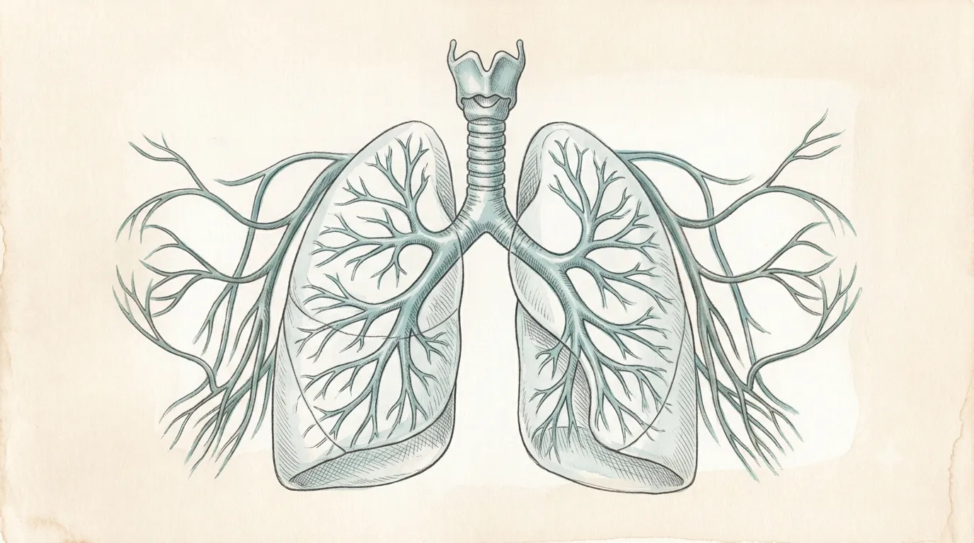
The right lung carries three lobes and the left only two, leaving a notch for the heart.
Respiratory System
Lungs
PulmonesThe breath of life
Exchanges oxygen for carbon dioxide
Paired organs that draw in air and trade oxygen for carbon dioxide across a vast, delicate surface.
- TracheaCarries air to the lungs
- CarinaThe ridge where the trachea forks in two
- Right LungThree lobes
- Left LungTwo lobes, room for the heart
- Lung ApexRises above the first rib, into the base of the neck
- BronchusBranching airway
- HilumThe root — vessels, airway, and nerves enter here
- Oblique FissureDeep cleft separating the lobes
- Cardiac NotchScoop in the left lung where the heart sits
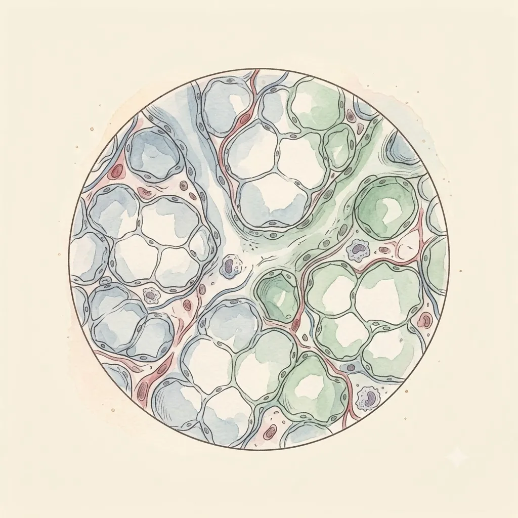 Under the lens
- Lung BaseRests on the diaphragm
- DiaphragmThe muscle that actually does the breathing
- Horizontal FissureOn the right lung only — the shelf that cuts the superior lobe off from the middle one
- Middle LobeThe right lung's third lobe, small and set at the front; the left has no counterpart to it
- LingulaThe tongue of left lung hanging under the cardiac notch — the middle lobe's opposite number in everything but name
- Left Main BronchusLonger and more horizontal than the right one, which is why an inhaled object usually goes down the other side
- Costodiaphragmatic RecessThe empty pleural gutter below the lung's own edge — where fluid settles, and where a drain is aimed
- Costal PleuraThe parietal layer against the ribs; somatic nerves supply it, and that is what makes pleurisy sharp
Structure
- Parietal pleuraLines the chest wall — the layer that hurts in pleurisy. It is named for whatever it covers: costal over the ribs, mediastinal, diaphragmatic, and a cervical cupula over the apex. Below the lung's own lower border it runs on for two more ribs, and that empty gutter is where fluid settles first
- Pleural spaceA film of fluid a few microns thick, holding the lung against the wall by surface tension
- Visceral pleuraWraps the lung itself and has no pain fibres
- Alveolar wallWhere exchange happens — under a micron between air and blood
- Surfactant filmDetergent-like coating stopping the smallest alveoli collapsing
- Sits
- Either side of the heart, within the ribcage
- Size
- Each about 25 cm tall
- Weight
- About 1 kg for the pair
The lesson
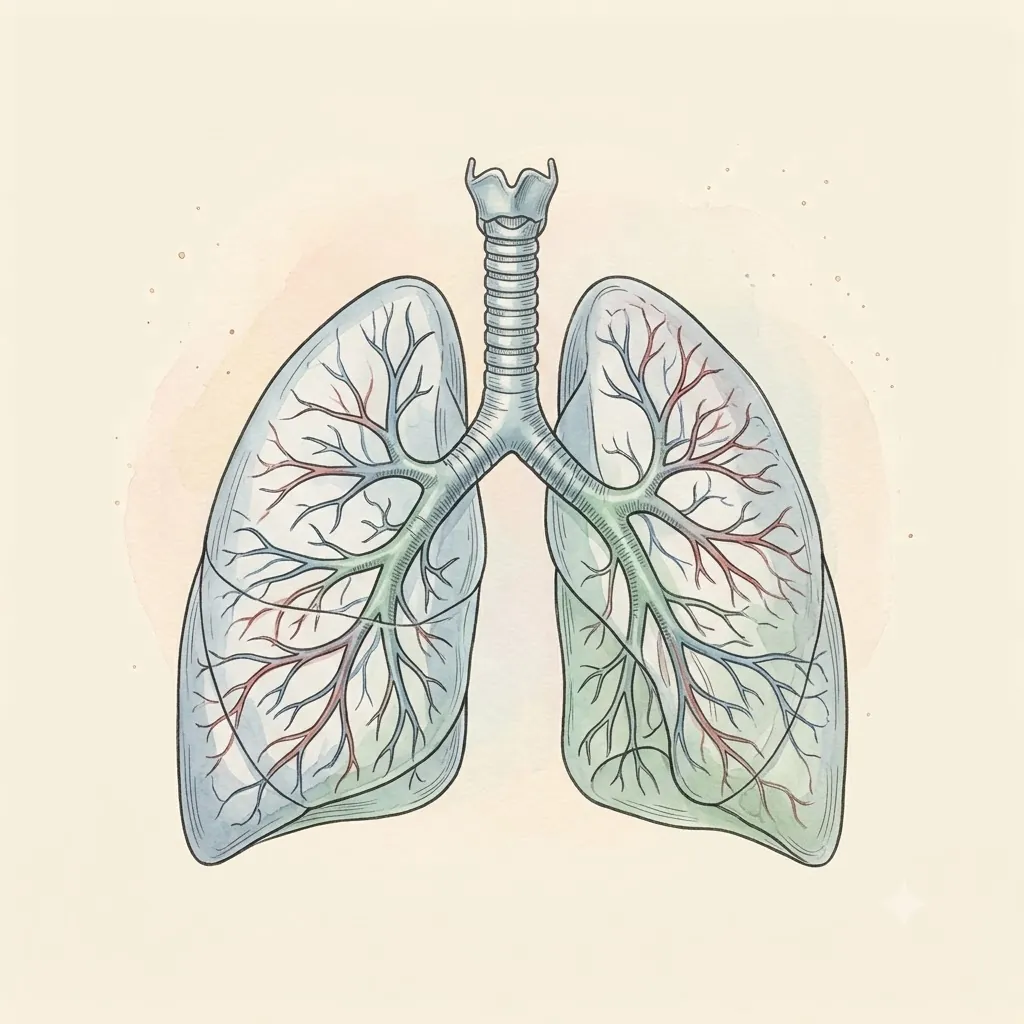
Follow one breath from the trachea, forking at the carina into a tree of narrowing airways, down to alveoli thinner than a sheet of paper. The right lung's three lobes and the left's two are not symmetrical by accident — the left gives up room for the heart's cardiac notch.
Twenty-odd divisions to reach the air
The airway begins as one tube and halves about twenty-three times before it ends. Each split makes the tubes narrower but doubles how many there are, so the total cross-section grows steeply as you descend.
Air rushes through the trachea and nearly stops in the smallest branches — which is exactly right, because exchange needs contact time, not speed.
The first sixteen or so generations exchange nothing at all; they only conduct. That volume is dead space — air that goes in and comes out without ever meeting blood. It is why shallow, rapid breathing ventilates so badly: a small breath may barely clear the tubing before it is exhaled again.
Remember
- Branching multiplies cross-section, so airflow slows toward the alveoli
- The conducting airways are dead space — no exchange happens there
- Shallow fast breaths waste a far larger fraction on dead space than slow deep ones
Nothing pumps the oxygen across
No carrier moves oxygen through the alveolar wall. It diffuses, driven only by the difference in partial pressure between air and blood, over a barrier well under a thousandth of a millimetre thick.
The design problem is therefore purely geometric: maximise the area, minimise the thickness, keep the two sides in contact long enough.
A red cell spends under a second in an alveolar capillary and is fully loaded in roughly a third of that, leaving a wide margin.
Disease eats the margin before it touches the result — thicken the wall or flood the space and the reserve goes first, which is why breathlessness shows up on exertion long before anything looks wrong at rest.
Remember
- Exchange is passive diffusion down a partial-pressure gradient
- Rate depends on area, barrier thickness and contact time
- Reserve disappears before resting gas levels move — exertion exposes it first
The lungs are inflated, not self-inflating
Lung tissue contains no muscle able to expand it. The diaphragm and chest wall enlarge the cavity, pressure inside drops below atmospheric, and air is pushed in from outside. Breathing in is active work; breathing out at rest is mostly elastic recoil being allowed to happen.
What couples the lung to the chest wall is a fluid seal, not an attachment — surface tension holding two wet surfaces together, the way two wet glass slides resist being pulled apart yet slide freely across each other.
Let air into that space and the lung springs away from the wall and collapses, while remaining entirely undamaged itself.
Remember
- Inspiration is active; quiet expiration is passive recoil
- The pleural fluid seal is what makes the lung follow the chest wall
- Air in the pleural space collapses a healthy lung — the seal failed, not the tissue
Clinical use
Moves around 11,000 L of air
Alveoli fold a tennis-court-sized exchange surface into the chest.
Always read a plate alongside the clinical picture.