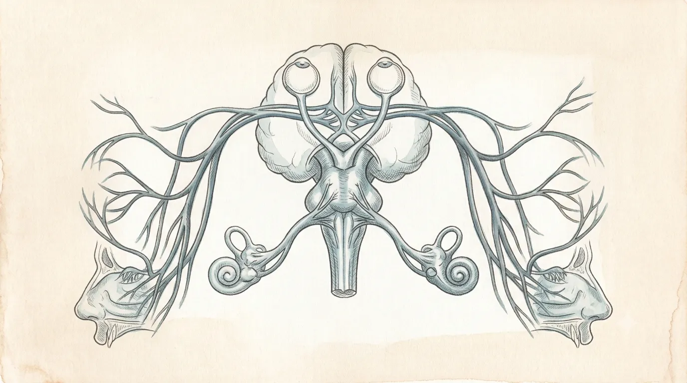
The cornea carries no blood vessels at all; it takes oxygen directly from the air.
Sensory System
Eye
OculusA window made of light
Captures and focuses light
A precision sensory organ that converts focused light into neural signals interpreted as vision.
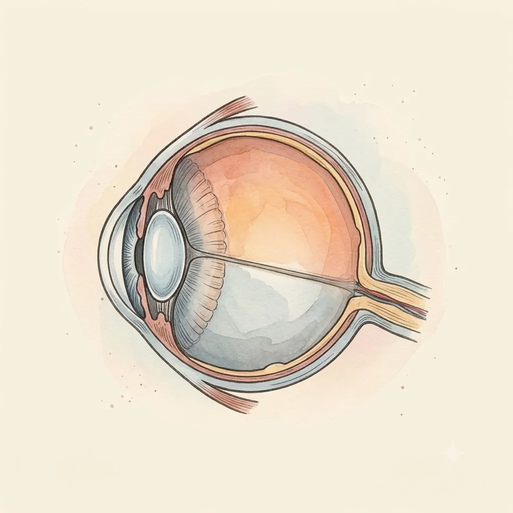
- CorneaClear focusing surface
- PupilNot a structure — the hole the iris leaves
- IrisControls light entry
- LensChanges shape to focus near and far
- Ciliary BodyMuscle that reshapes the lens and makes aqueous fluid
- ScleraThe tough white shell holding the eye's shape
- RetinaLight-sensing layer — brain tissue, not eye tissue
- MaculaTiny central patch carrying all fine detail vision
 Under the lens
- Optic NerveCarries visual signals
- Zonular FibresSuspending threads that hold the lens and keep it pulled flat
- ChoroidVascular sheet feeding the outer retina from behind
- LimbusCorneoscleral rim — the drainage angle lies just inside it
- Optic DiscWhere the fibres leave through the sieve in the sclera — the blind spot
- Lateral RectusTurns the eye outward — the abducens nerve's only muscle
- Medial RectusTurns the eye inward — the largest of the four recti
- Superior ObliqueRuns through a pulley in the orbit's roof — the trochlear nerve's only muscle
Structure
- Cornea & scleraOuter coat — transparent at the front, opaque white around the rest. The cornea is five layers deep: an epithelium that renews in days, Bowman's layer, a stroma whose collagen is spaced evenly enough that light crosses without scattering, Descemet's membrane, and an endothelium that pumps water back out and never divides, so its cells are simply spent across a lifetime. Behind it the sclera is the same collagen laid down at random, thinnest where the recti insert, and drilled at the back into the sieve of the lamina cribrosa the nerve fibres pass through.
- UveaVascular middle layer: iris, ciliary body, and choroid
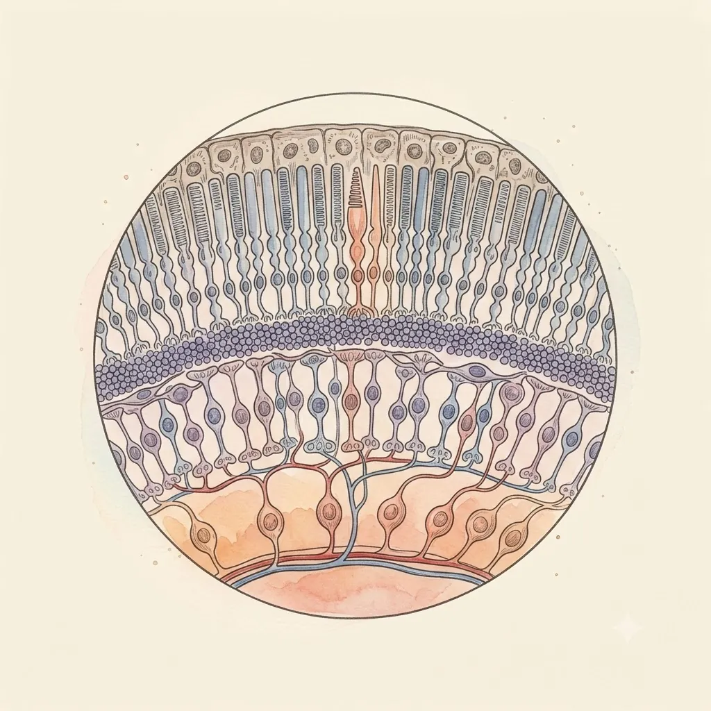
- RetinaNeural layer where photons become signals
- Pigment epitheliumBlack backing that absorbs stray light and recycles photopigment
- VitreousClear gel filling the globe and holding its shape
- Sits
- Within the bony orbit
- Size
- About 24 mm across
- Weight
- Around 7.5 g
The lesson
Light crosses several structures before it becomes a signal: the cornea bends it, the iris limits it, the lens focuses it, the vitreous carries it, and the retina — itself displaced brain tissue — converts it. Notice how little of the eye is devoted to focusing compared to sensing; most of its volume is simply getting light cleanly to the back.
Most of the bending happens before the lens
The lens gets the credit, but around two thirds of the eye’s focusing power belongs to the cornea, simply because that is where light crosses the biggest jump in refractive index — from air into tissue. The cornea’s contribution is fixed. The lens supplies the adjustable remainder.
Accommodation then works the opposite way round from intuition. The lens is elastic and would sit rounded if left alone; the fibres suspending it are under tension and pull it flat.
Contracting the ciliary muscle slackens that tension and lets the lens bulge for near work — so the relaxed eye is focused far away, and close focus is the effortful state.
The lens stiffens steadily with age until it can no longer round up, which is why reading glasses arrive more or less on schedule.
Remember
- Cornea supplies about two thirds of the refractive power, and cannot change
- Ciliary contraction slackens the suspending fibres and lets the lens round up
- Presbyopia is the lens stiffening — mechanical, not neural
Installed backwards, and it shows
The retina faces away from the light. Photoreceptors sit at the very back, so light has to cross the nerve layers and blood vessels lying in front of them before it reaches the cells that detect it. Vision is excellent despite this arrangement, not because of it.
Two consequences follow directly. Where the nerve fibres gather to leave the eye there can be no receptors at all, leaving a genuine hole in the visual field that goes unnoticed only because the brain fills it in.
And at the point of sharpest vision the overlying layers are swept aside into a small pit, so light there reaches the receptors unobstructed. All your fine detail comes from a patch roughly the width of a letter on this page.
Remember
- Light crosses the wiring before reaching the photoreceptors
- The optic disc has no receptors — a real blind spot the brain conceals
- Sharp vision comes from a tiny central pit where the layers are cleared away
An eye is a pressurised vessel
The optics only hold if the shape holds, and the shape is maintained by internal pressure. Fluid is produced continuously behind the iris, flows forward through the pupil, and drains at the angle where iris meets cornea. Production runs steadily; the drain is the variable part.
That makes the drainage angle the weak point. If outflow falls behind production, pressure rises, and what gives way is not the tough outer wall but the optic nerve fibres where they leave the eye.
Because the fibres serving the periphery go first, the loss starts at the edges of the visual field and reaches the centre last — often after years in which nothing was noticed and nothing hurt.
Remember
- Aqueous humour is made continuously and drains at the iridocorneal angle
- Internal pressure is what holds the eye’s shape, and therefore its focus
- Raised pressure kills nerve fibres from the periphery inward, painlessly
Clinical use
Makes thousands of tiny movements
The retina is an extension of the central nervous system.
Always read a plate alongside the clinical picture.