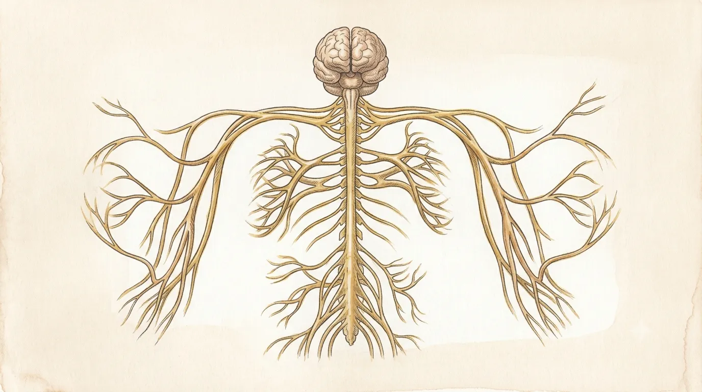
It has no pain receptors of its own — a headache is felt in the tissues around it.
Nervous System
Brain
EncephalonThe universe within
Processes and coordinates signals
The body’s command center, integrating sensation, memory, emotion, and precise movement.
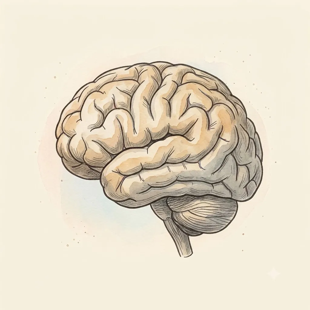
- Frontal LobePlanning & movement
- Motor CortexIssues the commands for voluntary movement
- Somatosensory CortexReceives touch from the whole body, mapped in order
- Parietal LobeSensory integration
- Temporal LobeMemory & hearing
- Occipital LobeVision — at the back, furthest from the eyes
- Broca’s AreaProduces speech; damage here leaves comprehension intact. It sits in the left inferior frontal gyrus in almost everyone, whichever hand they write with
- Wernicke’s AreaUnderstands speech, at the back of the left superior temporal gyrus — damage here leaves fluent, well-formed sentences that mean nothing
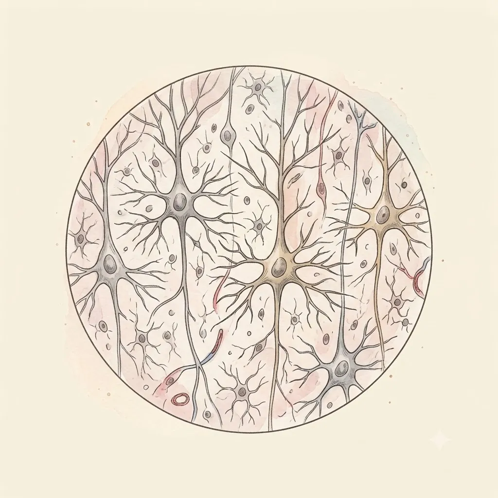 Under the lens
- Central SulcusThe groove that separates the motor strip in front from the sensory strip behind — the one landmark the whole cortex is oriented from
- Lateral SulcusThe deep cleft prising the temporal lobe away from the frontal; the middle cerebral artery runs up inside it, and the insula is buried at its floor
- Longitudinal FissureThe deep groove dividing left and right hemispheres, with the falx cerebri hanging down into it
- CerebellumBalance & coordination, tucked under the tentorium cerebelli behind the brainstem
- VermisThe cerebellum's midline ridge, joining the two hemispheres; it keeps the trunk upright while the hemispheres handle the limbs
- PonsThe brainstem's forward bulge, a bridge of fibres crossing into the cerebellum — and where the trigeminal nerve leaves
- BrainstemBreathing, heartbeat, consciousness — the non-negotiables
- Pituitary GlandHangs from the hypothalamus into its own saddle of bone, close enough to the optic chiasm that a growing tumour takes the outer half of each visual field
Structure
- Scalp & skullThe outer armour — bone up to 7 mm thick over the vault
- Dura materTough leathery sheet, the outermost of three membranes — two layers that split apart to carry the venous sinuses and fold inward as the falx cerebri and the tentorium cerebelli, partitioning the cavity
- Arachnoid & CSFWeb-like layer over a fluid cushion the brain effectively floats in
- Pia materDelicate film following every fold of the surface
- Grey matterCell bodies — the cortex, roughly 2–4 mm deep and heavily folded
- White matterMyelinated axons, the wiring that connects regions to each other
- Sits
- Protected within the skull
- Size
- Roughly two clenched fists
- Weight
- 1.3–1.4 kg
The lesson
Peel through the meninges — dura, arachnoid, pia — before reaching the folded grey matter where signals begin. Each lobe answers a different question: the frontal lobe plans and moves, the parietal integrates sensation, the temporal handles memory and hearing, and the occipital renders vision at the very back, farthest from the eyes it serves.
Grey thinks, white connects
The cortex is thin — a couple of millimetres of cell bodies — and almost everything beneath it is cable. That ratio is the design: a modest sheet of processing surface, and an enormous investment in wiring it to itself.
Folding is what buys the surface. Spread flat, the cortex would cover far more area than a skull can enclose, so it crumples, and about two thirds of it ends up hidden down in the grooves rather than exposed on the visible bulges.
Its thickness barely varies from place to place; what varies is what each patch is connected to.
Remember
- Grey matter is cell bodies, white matter the myelinated wiring between them
- Most cortex is buried in the folds, not on the visible surface
- Regions differ by their connections far more than by their structure
Territory, not talent
A lobe is not a faculty. Calling the occipital lobe “vision” names where visual signals arrive first, not a module that sees. The lobes are named after the skull bones above them, and nearly every real task recruits several at once.
What the division does capture is order. Signals land in a primary area, then spread outward into association cortex where they are combined with memory and context.
Damage to a primary area removes a raw channel; damage further out leaves the channel intact and the meaning gone — which is how someone can see a face perfectly clearly and not recognise it.
Remember
- Lobes take their names from the overlying bones, not their jobs
- Primary areas receive; association areas interpret
- Losing interpretation rather than input produces the strangest deficits
An organ that cannot wait
The brain runs an expensive metabolism continuously and stores almost no fuel of its own. It cannot power down the parts it is not using either: the gap between hard concentration and idle rest is a few percent, not the doubling intuition suggests.
That is why interrupting supply is so unforgiving. Muscle tolerates minutes of poor perfusion; cortical neurons start failing within seconds and die within minutes.
The circulation compensates with autoregulation — brain blood flow is held nearly constant across a wide range of blood pressures, a protection few other organs bother with.
Remember
- Almost no stored fuel, so supply must be continuous rather than merely adequate
- Effort changes consumption by a few percent, not by multiples
- Blood flow is autoregulated to stay near-constant as pressure swings
Clinical use
Uses about 20% of the body’s energy
Billions of neurons communicate through electrical and chemical signals.
Always read a plate alongside the clinical picture.