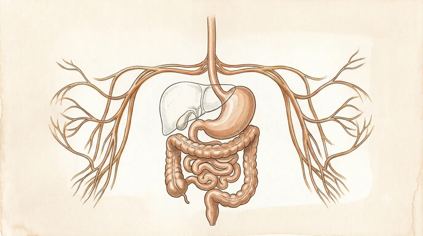
It is the only human organ that can regrow to full size from a fraction of itself.
Digestive System
Liver
HeparThe quiet alchemist
Metabolism, detoxification & bile
A remarkable metabolic organ that filters blood, processes nutrients, and produces bile.
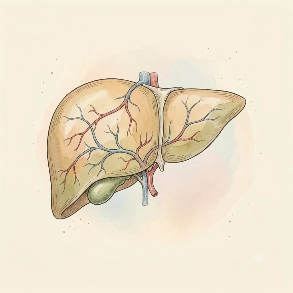
- Right LobeLargest hepatic lobe
- Left LobeCrosses the midline
- Falciform LigamentThe fold marking the surface division of the lobes
- Caudate LobeTucked behind, with its own blood supply
- Quadrate LobeOn the underside, beside the gallbladder
- Portal VeinNutrient-rich inflow
- Hepatic VeinsOutflow, straight into the vena cava
- GallbladderStores and concentrates bile until fat arrives
 Under the lens
- Common Bile DuctCarries bile down to the duodenum
- Diaphragmatic SurfaceThe smooth dome moulded to the diaphragm, with the right lung and pleura just above it
- Bare AreaThe one patch with no peritoneum over it, fused directly to the diaphragm
- Groove for the Vena CavaThe vein is bedded into the liver's back, and the hepatic veins join it here
- Ligamentum VenosumThe closed ductus venosus, lying in its fissure beside the caudate lobe
- Porta HepatisThe gate on the underside — vein, artery and ducts all cross here
- Proper Hepatic ArteryA quarter of the inflow, but nearly all the blood the bile ducts get
- Round LigamentThe fetal umbilical vein, closed off in the free edge of the falciform
Structure
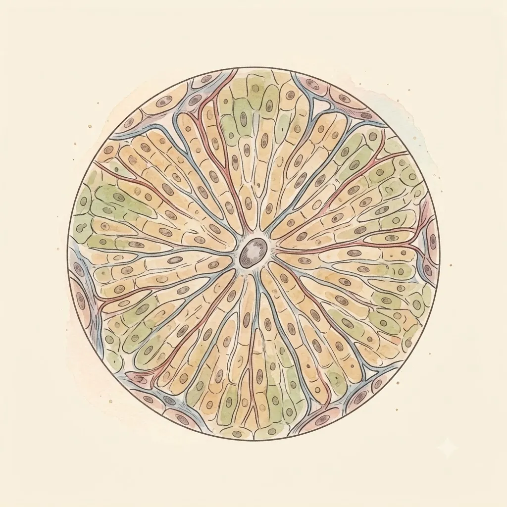
- Glisson's capsuleFibrous covering — the only part with pain fibres. It sleeves the vessels at the porta and follows them all the way in, so every portal tract is wrapped in it; stretch it quickly, as a swollen or congested liver does, and the ache appears under the right ribs.
- LobulesHexagonal working units, each built around a central vein
- HepatocytesThe workhorse cells, arranged in plates one or two cells thick
- SinusoidsLeaky capillaries letting blood bathe hepatocytes directly
- Bile canaliculiTiny channels running counter to the blood, collecting bile
- Sits
- Upper right abdomen
- Size
- About the size of a football
- Weight
- 1.4–1.6 kg
The lesson
The liver hides its complexity in repetition: millions of near-identical lobules, each a hexagon built around a central vein, each doing the same quiet work of filtering, storing, and building. Blood arrives two ways — oxygen-rich from the hepatic artery, nutrient-rich from the portal vein — and leaves by a single route, carrying the results of both.
Why a second blood supply changes everything
Most organs take arterial blood and hand it back to a vein. The liver also receives the portal vein, already stripped of its oxygen but loaded with everything absorbed from the gut, and about three quarters of the liver’s blood arrives that way. Oxygen is the minority supply here.
The consequence is that the liver inspects the intestinal harvest before the rest of the body sees any of it, and can alter it in passing.
A swallowed drug is worked over here first, and the fraction that survives to reach the circulation is often small — which is why some drugs are useless taken by mouth and have to be injected, and why the same dose behaves differently by the two routes.
Remember
- Roughly three quarters portal (nutrient-rich), one quarter arterial (oxygen-rich)
- Everything absorbed from the gut passes the liver before reaching the body
- First-pass metabolism is why oral and injected doses are not interchangeable
One cell type, doing all of it
Nothing defines the liver the way a beat defines the heart.
The same cell banks glucose as glycogen and releases it hours later, assembles most of the plasma proteins including albumin and the clotting factors, and converts the ammonia left from broken-down protein into urea the kidneys can carry away.
Uniformity is the trick. Hepatocytes are largely interchangeable rather than divided up by task, so the organ scales by adding more of the same.
That is also why its failures are so quiet: a large share of the tissue can be gone before the remainder stops keeping up, and the first sign is usually a blood test rather than a symptom.
Remember
- Glycogen store, plasma-protein factory, and the body’s ammonia disposal
- Interchangeable cells mean the organ scales by bulk, not by specialisation
- Clotting factors are made here, so liver disease shows up as bleeding
Bile is a detergent and a bin
Bile does two unrelated jobs at once. As a detergent it breaks dietary fat into droplets small enough for enzymes to attack — it contains no digestive enzymes of its own, and without it fat passes through largely undigested.
As a disposal route it carries out what the kidneys cannot: substances too large or too fat-soluble to leave in urine, including the pigment left over from worn-out red cells.
That pigment is bilirubin, and following it through the plumbing explains most of what a jaundiced patient looks like.
When bile cannot get out — the liver unable to process it, or the duct blocked — bilirubin backs up into the blood and stains the skin and the whites of the eyes, the kidneys start dumping it so the urine darkens, and the stool loses the colour bile normally gives it. One molecule, three signs.
Remember
- Bile emulsifies fat; the enzymes that digest it come from elsewhere
- It is also the exit route for waste urine cannot carry
- Backed-up bilirubin explains yellow skin, dark urine and pale stool together
Clinical use
Performs more than 500 functions
It can regenerate a substantial portion of lost tissue.
Always read a plate alongside the clinical picture.