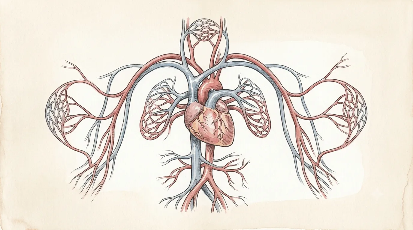
It beats roughly 2.5 billion times in an average lifetime, and starts before you are born.
Cardiovascular System
Heart
CorThe tireless pump
Circulates oxygenated blood
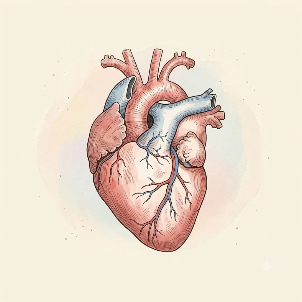
A muscular organ that pumps blood throughout the body, delivering oxygen and nutrients to every cell.
- AortaMain artery
- Pulmonary TrunkCarries venous blood to the lungs
- Superior Vena CavaReturns blood from head and arms
- Inferior Vena CavaReturns blood from the abdomen and legs
- Left AtriumReceives oxygenated blood
- Right AtriumReceives venous blood
- Sinoatrial NodeThe pacemaker — sets the rhythm itself
- Atrioventricular NodeThe only electrical door between atria and ventricles, and it deliberately delays every signal it passes
- Left VentriclePumps to the body
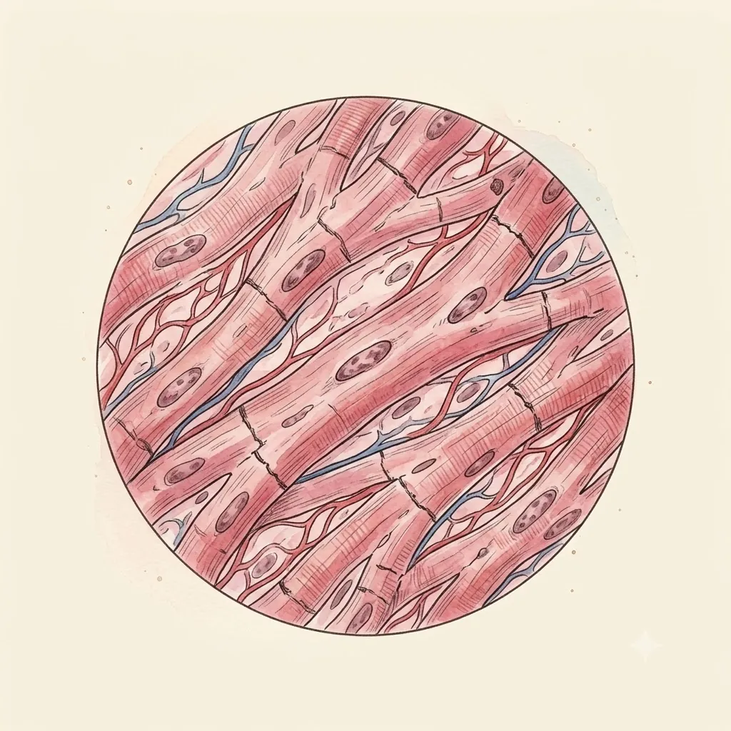 Under the lens
- Right VentriclePumps to the lungs
- Mitral ValvePrevents backflow
- Tricuspid ValveGuards the right atrium's outflow
- Aortic ValveThree cupped leaflets at the aorta's mouth; the coronary arteries leave from two of them
- Pulmonary ValveShuts the pulmonary trunk the instant the right ventricle relaxes — the second heart sound, right half
- Anterior Interventricular ArteryThe LAD, running the front groove between the ventricles; it feeds the septum and the left ventricle's front wall
- Right Coronary ArteryCircles in the right coronary sulcus and supplies the sinoatrial node in most hearts
- Coronary SinusThe heart's own vein, collecting spent blood at the back and emptying straight into the right atrium
- ApexThe tip, where the beat is felt on the chest wall
Structure
- PericardiumTough outer sac holding a teaspoon or two of fluid so the heart can move without friction — fill it fast and the heart cannot fill at all
- EpicardiumOuter surface of the heart itself, carrying the coronary vessels and fat
- MyocardiumThe thick muscle that does the pumping — far thicker on the left
- EndocardiumSmooth inner lining, continuous with the vessels, keeping blood from clotting
- Sits
- Behind the sternum, slightly left
- Size
- About the size of your fist
- Weight
- 250–350 g
The lesson
Start at the surface and work inward: the pericardium's slick sac lets the heart move without friction, the myocardium does the actual pumping, and four chambers hand blood off in one direction only, kept from flowing backward by the mitral and tricuspid valves. Trace a single drop from the vena cava through the right heart to the lungs, back through the left heart, and out the aorta — the whole circuit in four beats.
Blood takes two laps, not one
The heart is not one pump serving one loop. It is two pumps in series, each serving a different circuit: the right side sends blood the short distance to the lungs and back, the left drives it around the entire rest of the body.
Both eject the same volume with every beat, and they must — any lasting mismatch would drain one circuit into the other within minutes.
That is why the two halves are built so differently while doing the same job. The pulmonary circuit is short and its vessels offer little resistance, so the right ventricle works against roughly a fifth of the pressure the left does. Wall thickness follows the pressure, not the volume.
Remember
- Right heart drives the pulmonary circuit, left heart the systemic
- Both ventricles move equal volumes; only the pressures differ
- Systemic pressure is several times pulmonary — hence the thicker left wall
Valves are what make it a pump
A muscular chamber that squeezes is not yet a pump. What makes it one is that blood can only leave in the direction the valves permit: squeeze a chamber whose exit is open and whose entrance is shut, and everything inside goes forward.
The valves are entirely passive — no muscle opens or closes them. They move on pressure difference alone. When pressure in a ventricle rises above the atrium behind it, the mitral and tricuspid are pushed shut; when it climbs past the artery ahead, the aortic and pulmonary are pushed open.
The two heart sounds are those two closures, in that order.
Remember
- Pressure difference alone opens and shuts every valve
- First sound = mitral and tricuspid closing; second = aortic and pulmonary
- A leaking valve wastes work — blood sent forward comes back
The rhythm starts inside the muscle
Cut every nerve to the heart and it keeps beating. The signal starts in the sinoatrial node, a patch of cells that leak ions steadily until they reach threshold and fire. It is a clock, not a relay. Nerves only tell it to hurry or to slow.
The wave then spreads through muscle that is electrically joined cell to cell, so a chamber contracts as a unit rather than fibre by fibre.
A deliberate pause at the atrioventricular node holds the signal for a fraction of a second, and that pause is what lets the atria finish emptying before the ventricles squeeze. Remove it and the two would contract together, moving far less blood per beat.
Remember
- The sinoatrial node fires on its own; nerves adjust the rate, not the beat
- Electrically coupled muscle contracts as a single unit
- The atrioventricular delay is functional: atria empty first
Clinical use
Beats about 100,000 times
Its electrical rhythm coordinates every heartbeat.
Always read a plate alongside the clinical picture.