The uterine tube does not touch the ovary. The egg is released into the abdominal cavity and swept up by the fimbriae — a gap of a few millimetres that the whole tract is built around.
All modules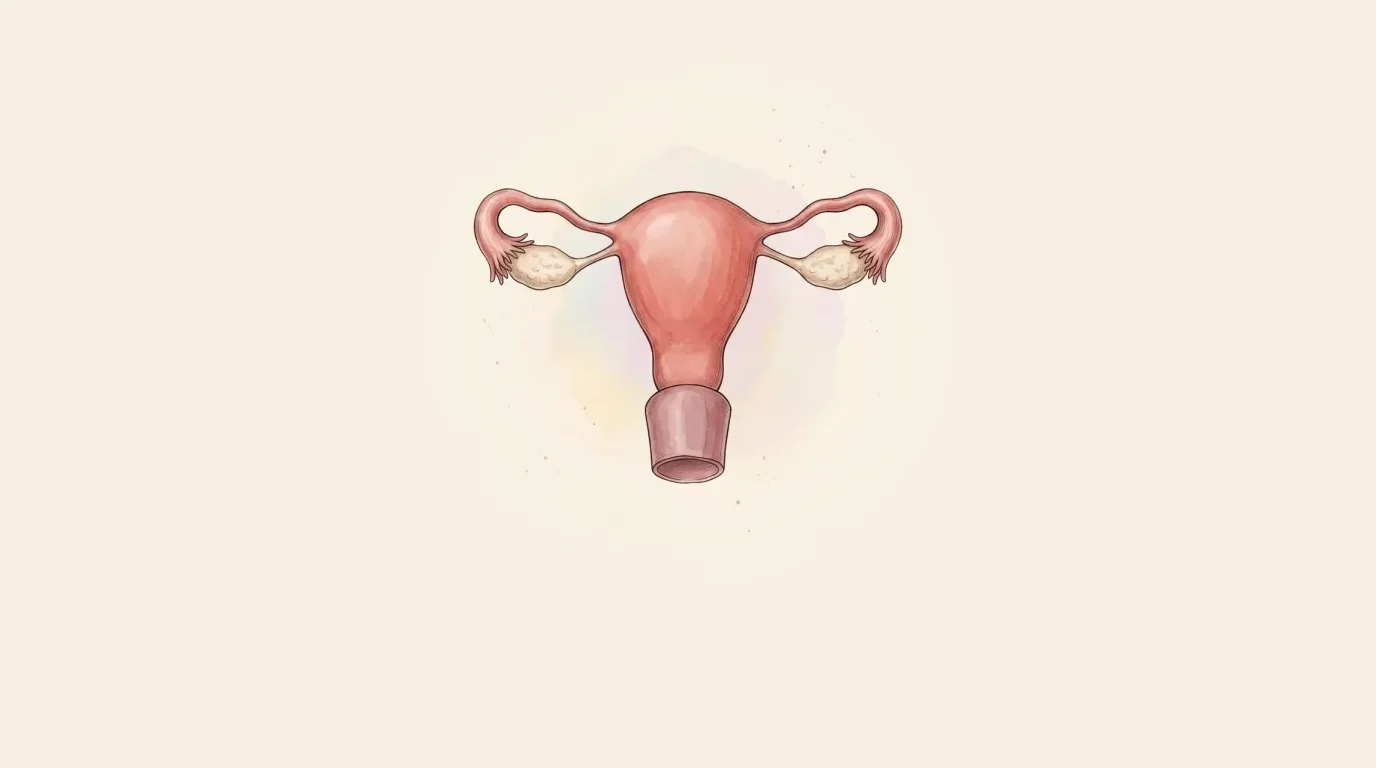 Reproductive System
Female genital tract: the lesson
Structure and story of the female genital tract, layer by layer.
Reproductive System
Female genital tract
Organa genitalia feminina internaThe one passage open at both ends
Makes eggs and hormones, and carries a pregnancy
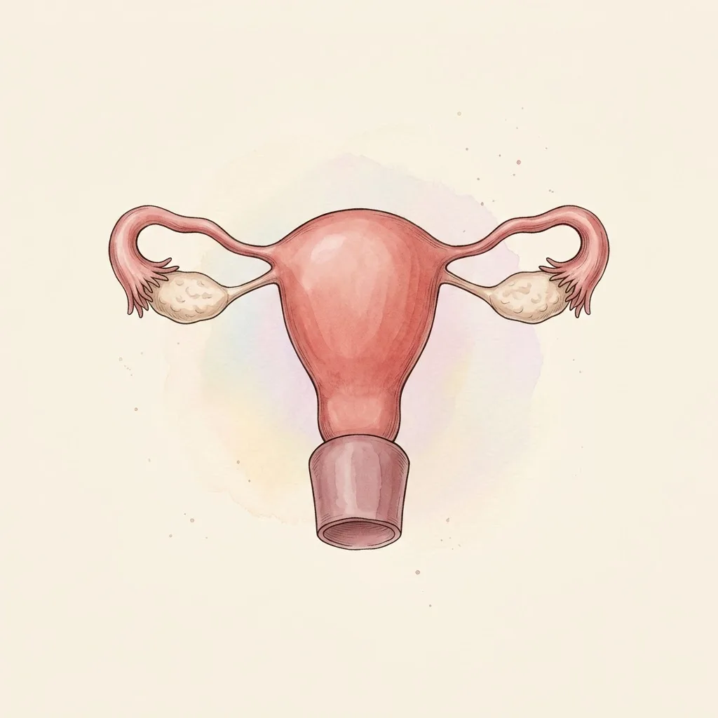
Ovary, tube, uterus and vagina — a tract that makes an egg, catches it in open abdomen, and holds what may follow.
- AmpullaThe wide outer third, where fertilisation happens
- Tubal isthmusThe narrow inner stretch — about 1 mm across
- CornuThe horn where the tube leaves the uterine cavity
- FimbriaeThe fringe that sweeps the ovary's surface — the tube's only contact with the egg it collects
- Ovarian ligamentAnchors the ovary to the uterine horn, under the tube
- OvaryGamete and hormone in one organ, and the only one left uncovered by peritoneum
 Under the lens
- FundusThe dome above the tubes — the height measured in pregnancy
- IsthmusThe waist where the body bends forward over the cervix
- CervixThe neck that stays shut for nine months and opens in hours
- VaginaA muscular canal whose walls rest against each other until something opens them
Not visible on this view
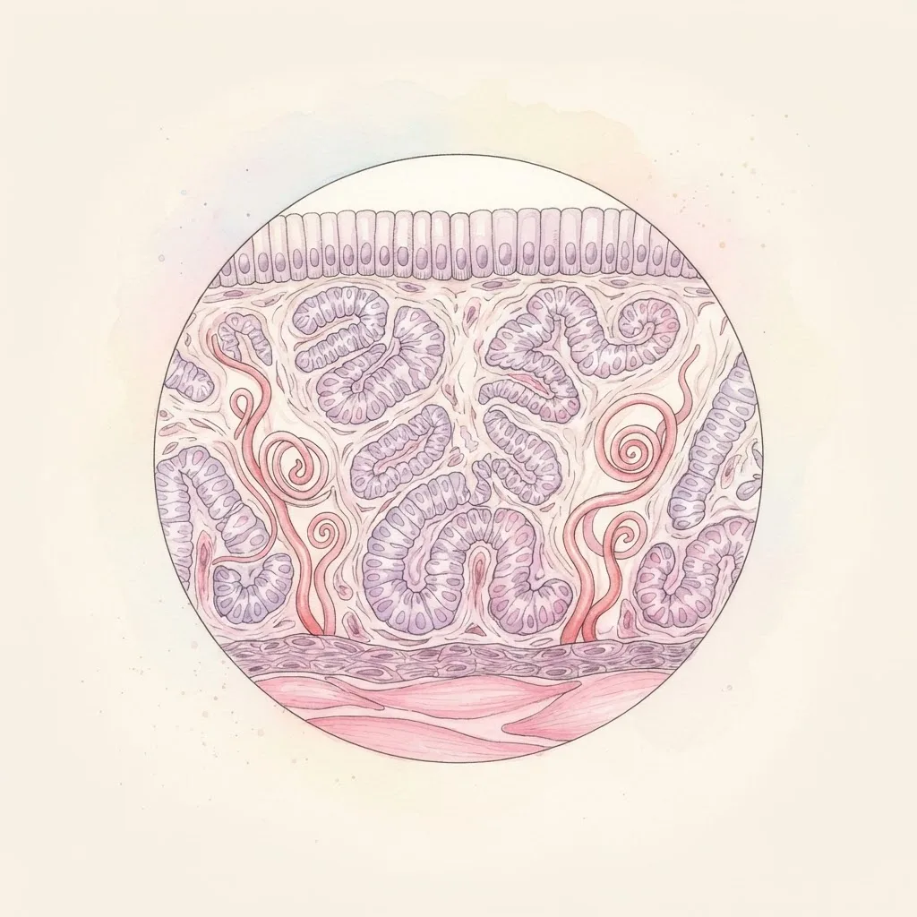
- EndometriumThe lining inside this wall: rebuilt and shed with each cycle, and the tissue a pregnancy implants into
- MyometriumThree interwoven layers of smooth muscle — they contract around the vessels running through them, which is what stops bleeding after birth
- External osThe opening into the vagina, and the transformation zone around it where cervical cancer starts
- FornixThe gutter around the cervix — the posterior one is where the pouch behind the uterus is felt
Structure
- PerimetriumThe peritoneal covering, continuous with the broad ligament on each side
- MyometriumThe bulk of the wall: smooth muscle in three directions, so contraction squeezes rather than shortens
- Functional layerGrown, held, and shed each cycle — the part that becomes the site of implantation or the menstrual loss
- Basal layerKept through every cycle; the source the functional layer regrows from, and irreplaceable once scarred
- Sits
- In the pelvis, between the bladder in front and the rectum behind
- Size
- Uterus about 7.5 cm long; tube about 10 cm
- Weight
- Uterus 60–80 g, doubling in the years of use
The lesson
This tract is arranged around one unusual fact: it is open. The vagina leads to the cervix, the cervix to the uterus, the uterus to the tubes, and the tubes end in an open funnel in the peritoneal cavity. Everything else the tract does — how it catches an egg, how it is examined, how infection travels through it — follows from that opening.
A tube that ends in nothing
The uterine tube does not attach to the ovary. It ends in a funnel of fingers that lies against it, and an egg leaving the ovary crosses a real gap of peritoneal cavity before the fimbriae sweep it in.
That is the only place in the body where the outside world connects to the abdomen, and the tract's whole clinical behaviour comes from it.
Read forwards, it explains how an egg is collected at all: the fimbriae move over the ovary's surface at ovulation, and the ciliated lining beats inward the whole length of the tube.
Read backwards, it explains what else travels that way — an infection at the cervix reaching the tubes and then the peritoneum, and blood from a ruptured tube filling the pouch behind the uterus rather than staying where it started.
Remember
- The egg crosses open abdomen between ovary and tube; nothing is joined
- It is the body's only route from outside to the peritoneal cavity
- Infection ascends the same path an egg descends
The only tissue that is meant to be lost
The endometrium is rebuilt and shed roughly four hundred times in a life, and it is the only tissue in the body whose normal state includes being destroyed on schedule.
It does it in two layers: a deep basal layer that stays, and a functional layer above it that grows under oestrogen, is held under progesterone and is lost when progesterone falls. What is shed is the functional layer only — the basal layer is the seed the next one grows from.
That two-layer arrangement is why the cycle is readable and why it fails in recognisable ways.
Bleeding is the withdrawal of hormone support, not an event in its own right, so anything that keeps progesterone high stops it and anything that leaves oestrogen unopposed lets the lining thicken until it outgrows its blood supply and bleeds unpredictably.
And because the basal layer is what regenerates, damaging it — by aggressive instrumentation or infection — costs the lining permanently.
Remember
- A basal layer stays; a functional layer above it is grown and shed
- Bleeding is hormone withdrawal, not a scheduled rupture
- Unopposed oestrogen thickens the lining; a damaged basal layer cannot rebuild it
Where fertilisation actually happens
Fertilisation does not happen in the uterus.
It happens in the ampulla, the wide outer third of the tube, within a day of ovulation, and the embryo then spends three or four days travelling down while it divides — arriving at the uterus as a blastocyst, at exactly the moment the lining is ready to accept it.
The timing is the point: the tube is not a corridor, it is a delay line.
This is why tubal disease is a fertility problem rather than a pain problem, and why an ectopic pregnancy is a tubal event.
If cilia are destroyed or the lumen is scarred — most often by past infection — the embryo cannot complete the journey, implants where it stopped, and grows into a wall that has no capacity to hold it.
The tube then bleeds into the abdomen, which is why a positive pregnancy test with pelvic pain is treated as an emergency until a scan shows the pregnancy inside the uterus.
Remember
- Fertilisation is in the ampulla; the uterus receives a blastocyst days later
- The tube's job is timing as much as transport
- A scarred tube traps the embryo — the anatomy of an ectopic pregnancy
Clinical use
About a thousand follicles are lost each month, one of them ovulated
The tract is the only route from the outside world to the peritoneal cavity, which is why infection here does not stay local.
Always read a plate alongside the clinical picture.