
Revision sheet
The anatomical position, planes and axes
The reference posture every description in the course silently assumes, and the three planes cut through it.
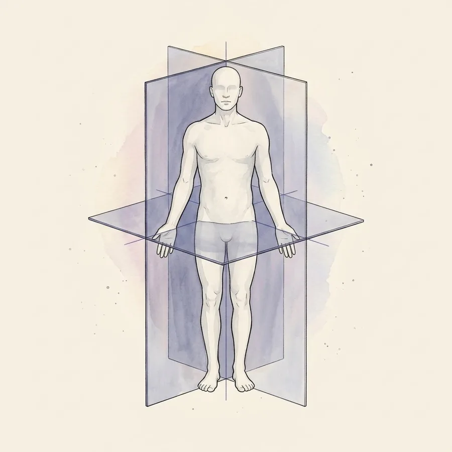
The position every description assumes
Anatomy describes a body in one fixed posture, and only that one: standing upright, feet together and flat on the ground, head and eyes facing forward, arms hanging by the sides with the palms turned forward.
Every term you will meet this year — anterior, superior, medial — is measured against that figure and nothing else.
The convention exists because the body moves. If you describe the thumb as "the outer finger", the sentence stops being true the moment the hand rotates. Anchor the description to the anatomical position and the thumb is lateral for good, whatever the arm happens to be doing when you examine it.
- Palms forward is the detail most often forgotten — it is what makes the radius lateral and the ulna medial.
- Left and right always mean the patient's left and right, never yours.
- A description is only valid against the anatomical position, never against how the body is lying in front of you.
Three planes, three ways to cut
A sagittal plane runs front to back and divides the body into left and right parts. The one passing exactly through the midline is the median (mid-sagittal) plane; any parallel cut off to one side is parasagittal.
A frontal (coronal) plane runs from side to side and separates anterior from posterior. A transverse (axial, horizontal) plane cuts across the body and separates superior from inferior — this is the plane of a routine CT slice.
- Sagittalleft / right
- Frontal (coronal)front / back
- Transverse (axial)top / bottom
The axes movement happens around
Each plane has an axis perpendicular to it, and a joint movement in a given plane turns around that axis. Flexion and extension happen in the sagittal plane, around a transverse (side-to-side) axis. Abduction and adduction happen in the frontal plane, around a sagittal (front-to-back) axis.
Rotation happens in the transverse plane, around a vertical (longitudinal) axis.
Learning the pairing once saves memorising every movement separately: name the plane a movement is drawn in and the axis follows, which is exactly how an examiner expects you to justify an answer.
- Sagittal plane, transverse axisflexion / extension
- Frontal plane, sagittal axisabduction / adduction
- Transverse plane, vertical axismedial / lateral rotation
Reading a slice the way radiology draws it
An axial CT or MRI slice is displayed by convention as though you were standing at the patient's feet looking up towards the head. The consequence trips up every first-year student: the patient's left side appears on the right of the image.
Coronal images are read as if facing the patient, so the same left–right inversion applies. Sagittal images have no inversion to worry about, but you must know which side the slice was taken from before you call a structure medial or lateral.
- Axial slice: viewed from the feet up — patient's left is on the right of the screen.
- Never call a finding "on the left" from an image without checking the side markers.